

Feeling a persistent ache, a subtle creak, or outright stiffness? These sensations often signal issues with your joints. Far more than mere connection points, joints are intricate biological marvels enabling movement, countless actions, and structural integrity. Understanding these complex structures is the crucial first step in addressing discomfort and ensuring long-term mobility.
Our bodies are masterpieces of engineering, and articulations are at their functional core. These sophisticated junctions link bones, ossicles, or other hard structures, constructing the skeletal system into a functional whole. They are meticulously built to facilitate a vast spectrum of movement, from precise surgeon’s hands to powerful athletic leaps. From protecting delicate organs to bearing immense loads, joints are indispensable to our daily existence.
As we delve deeper, we’ll explore the fundamental classifications and functions of these critical anatomical features. We’ll uncover how diverse joint structures dictate their capacity for movement. This in-depth research aims to empower you with a comprehensive understanding of your skeletal system, providing foundational knowledge to interpret stiffness, appreciate mobility, and proactively support your body’s incredible design.
1. **The Indispensable Role of Joints in Human Movement and Stability**Joints serve as the crucial linchpins of the human body, knitting together the skeletal system into a cohesive, functional whole. They are the sites where bones meet, forming fundamental connections for virtually every physical action we perform. Without these articulations, our bodies would be rigid, unmoving frameworks, utterly devoid of the flexibility and dynamic capabilities defining human life, making them indispensable for mobility.
Beyond enabling motion, joints are paramount for providing structural support and stability to the entire skeleton. They maintain posture, allowing us to stand upright and to sit or lie in various positions. This structural integrity is vital for balance, preventing us from toppling. Joints also bear significant loads, enduring daily activity stresses.
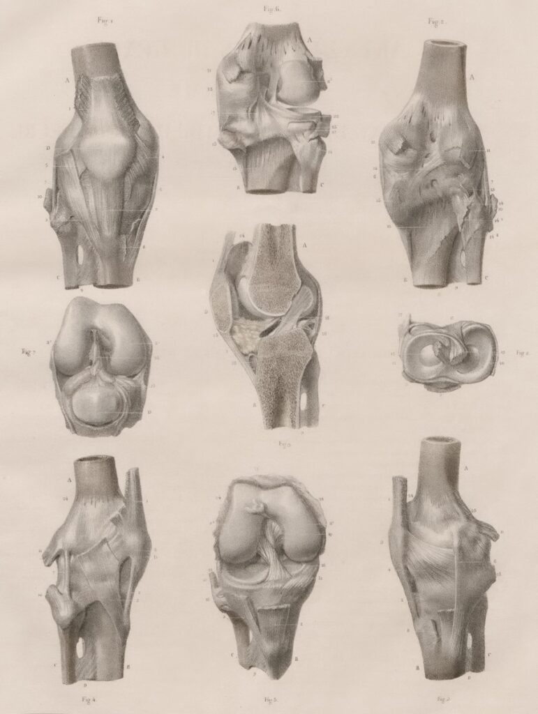
The range of movements facilitated by joints is astonishing. Consider the knee, elbow, and shoulder joints, described as “self-lubricating, almost frictionless, and able to withstand compression and maintain heavy loads while still executing smooth and precise movements.” This intricate design allows seamless execution of activities, underscoring their profound importance.
Read more about: Jennifer Aniston’s Odyssey to Enduring Strength: Unveiling Her “Strength Era” Through Pvolve

2. **Deconstructing the Joint: Essential Components and Connective Tissues**To truly appreciate joint functionality, understanding its constituent parts is essential. Joints are comprised of bones and specialized connective tissues holding them together. These tissues vary in composition and mechanical properties, directly influencing the joint’s flexibility, strength, and range of motion.
Key among these are cartilage, tendons, and ligaments. Cartilage, especially hyaline cartilage, often covers articulating bone surfaces, providing a smooth, low-friction medium for movement and shock absorption. Tendons are strong, fibrous cords connecting muscles to bones, transmitting force across the joint to enable movement.
Ligaments, also dense connective tissue, are crucial for joint stability, connecting bones to other bones. They act as robust biological ropes, preventing excessive or undesirable movements and helping to keep the joint properly aligned. Joints are also richly supplied with nerves, particularly unmyelinated fibers in capsules and ligaments, responsible for pain perception and providing proprioceptive feedback.

3. **Fibrous Joints: Unyielding Connections for Protection and Strength**Structural classification defines joints by their binding tissue. Fibrous joints are united by dense regular connective tissue, rich in collagen fibers. This robust connection typically results in joints with very little, if any, mobility.
These seemingly immobile connections play incredibly vital roles, especially where maximal protection or unyielding stability is needed. Functionally, most fibrous joints are synarthroses, permitting little to no movement. This immobility is a key design feature, exemplified by skull sutures protecting the brain.
Fibrous joints categorize into sutures, gomphoses, and syndesmoses. Sutures, in the cranium, tightly bind skull bones. Gomphoses are unique immobile joints between teeth and jaw sockets, held by the periodontal ligament. Syndesmoses, like the tibiofibular syndesmosis, offer slight mobility while maintaining long bone union, resisting divisive forces.

4. **Cartilaginous Joints: Flexible Foundations with Limited Mobility**Cartilaginous joints are another structural type, where bones are joined by cartilage. These joints offer a unique balance of limited movement, cushioning, and structural integrity. Functionally, most are amphiarthroses, permitting slight mobility, distinguishing them from immovable fibrous joints.
They divide into primary (synchondroses) and secondary (symphyses) types. Synchondroses, composed of hyaline cartilage, can be temporary (like the epiphyseal plate for bone lengthening) or permanent (like the first sternocostal joint, allowing no movement).
Symphyses, or secondary cartilaginous joints, incorporate thick, strong fibrocartilage known for resisting pulling and bending forces. Despite this strength, symphyses are amphiarthroses, allowing limited movement. Examples include the pubic symphysis, critical during childbirth, and the intervertebral symphysis (disk), which cushions between vertebrae, enabling spinal flexibility.

5. **Synovial Joints: The Apex of Mobility and Lubrication**When contemplating dynamic movement—arm swings, knee bends, head rotation—we are invariably considering synovial joints. These represent the pinnacle of human body mobility, functionally classified as diarthroses, or freely movable. Their sophisticated design permits a vast array of complex, essential daily actions.
The defining characteristic of a synovial joint is its unique “synovial cavity,” a fluid-filled space separating articulating bones. This cavity is encased by the articular capsule, a dense irregular connective tissue. Inside, the “synovial membrane” secretes “synovial fluid,” a remarkable lubricant reducing friction and facilitating smooth, precise movements.
Further enhancing this frictionless environment is “articular cartilage,” hyaline cartilage covering each bone’s articulating surface. This smooth layer is continuous with the synovial membrane, crucial for shock absorption and load distribution. These combined elements ensure synovial joints withstand compression and heavy loads while maintaining fluidity. Movement extent varies among subtypes, often limited by accessory ligaments.

6. **Hinge Joints: Precision Movement in a Single Plane**Among synovial joint classifications, the hinge joint exemplifies uniaxial articulation. Like a door hinge, it permits movement predominantly in a single plane. This design allows for a straightforward, powerful range of motion, primarily flexion (bending) and extension (straightening) along one axis.
Structurally, the convex end of one bone articulates with the concave edge of another. This interlocking shape restricts movement to a specific arc, ensuring stability and maximizing action efficiency. Its uniaxial nature means these robust joints allow significant force generation in primary directions but no rotation around other axes.
Common and critical examples include the elbow joint, enabling forearm flexion and extension. Similarly, the knee joint primarily functions as a hinge, allowing leg bending and straightening. The ankle and interphalangeal joints also fit this category, demonstrating the widespread importance of precise, single-plane movement for daily activities.

7. **Condyloid Joints: Biaxial Versatility for Intricate Actions**Expanding on synovial joint movements, the condyloid joint, or ellipsoid joint, offers greater versatility than uniaxial types. Characterized by articulation between a shallow depression and a rounded bone structure, this joint is notably biaxial. It permits movement across two distinct axes, allowing a more complex and varied range of motion.
Its biaxial nature enables four primary movements: flexion and extension (decreasing/increasing angles), and abduction and adduction (moving limbs away/towards midline). A key distinction from ball-and-socket joints is that condyloid joints do not allow full 360-degree rotation, despite some visual similarities.
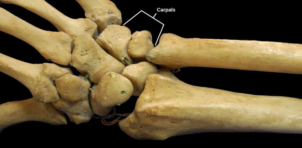
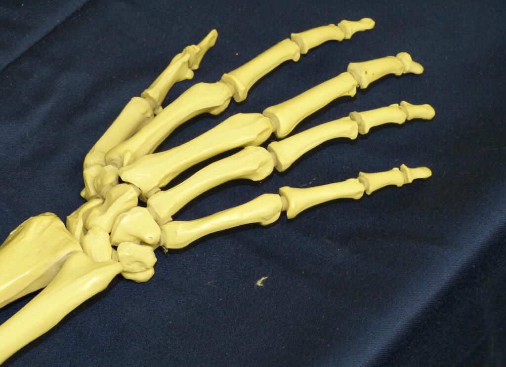
Excellent examples are found where intricate movements are essential, such as the hand and foot. The knuckles (distal metacarpals and proximal phalanges of the medial four fingers) are classic condyloid joints. The wrist (radiocarpal joint) and toe-to-foot joints also fit this classification, facilitating precise, coordinated actions for grasping, walking, and fine motor control.
Navigating the complexities of joint types and their biomechanics is crucial for understanding how our bodies move and what contributes to stiffness. Continuing our exploration, we turn our attention to additional synovial joint classifications and delve into the critical clinical considerations, neural networks, and muscular support systems that collectively maintain joint health and function. This comprehensive overview is designed to further empower you with the knowledge needed to appreciate the sophisticated design of your skeletal system and make informed decisions about your well-being.

8. **Saddle Joints: Unlocking the Power of the Opposable Thumb**Among the diverse array of synovial joints, the saddle joint stands out for its unique shape and the specialized movements it facilitates. This distinctive articulation occurs where two bones, each shaped like a saddle – concave in one direction and convex in another – meet. This interlocking design provides a balance of stability and impressive range of motion, classifying it as a biaxial joint.
Functionally, saddle joints allow for movement along two distinct axes. This means they enable a broader range of motion than condyloid joints, offering significant freedom. However, they do not permit full 360-degree rotation, distinguishing them from ball-and-socket joints in that specific aspect.
The most prominent example of a saddle joint in the human body is found at the base of the thumb, specifically where the trapezium bone articulates with the first metacarpal bone. This particular joint is pivotal, allowing the thumb to flex and extend parallel to the palm, and to abduct and adduct perpendicular to it. Such intricate movements are fundamental to the thumb’s opposable nature, a critical feature for grasping, manipulating objects, and performing complex hand motions. Without this joint’s unique capabilities, much of our dexterous hand function would be severely limited.
9. **Planar Joints: The Subtle Gliding for Flexibility and Load Distribution**Planar joints, often referred to as gliding joints, are characterized by their relatively simple structure, formed when two mostly flat or slightly curved bones articulate. These joints allow for one bone surface to slide or glide over another without significant rotation or angular movement. While seemingly subtle, these gliding motions are crucial for distributing forces and accommodating changes in joint position.
Functionally, planar joints are considered multiaxial, meaning they can permit movement in multiple planes. However, this movement is typically restricted by the strong surrounding ligaments that bind the bones together. The limited, sliding motion is essential for adaptability and load sharing across joint surfaces, preventing excessive stress on any single point.
Common examples of planar joints are found in areas requiring flexible yet stable connections. These include the intercarpal joints between the carpal bones in the wrist, and the intertarsal joints between the tarsal bones in the foot. These joints allow for slight movements that collectively contribute to the overall flexibility of the wrist and ankle. The acromioclavicular joint, connecting the acromion of the scapula and the clavicle, also falls into this category.
Furthermore, the joints between the articular processes of adjacent vertebrae in the spine are classified as planar joints. While the primary cushioning and flexibility between vertebrae are provided by cartilaginous intervertebral discs, these planar articulations facilitate the subtle gliding necessary for spinal movement. This arrangement allows the spine to twist, bend, and maintain its remarkable flexibility while also distributing compressive loads.
10. **Pivot Joints: Enabling Rotation and Head Movements**Pivot joints are specialized uniaxial synovial joints, designed to permit rotation around a single central axis. This unique design features the cylindrical end of one bone rotating within a ring formed by another bone and its encircling ligaments. This mechanical arrangement ensures controlled rotary movement while maintaining structural integrity.
The uniaxial nature of a pivot joint means that movement is limited to rotation in place, without the bone moving out of its original position along other axes. This precision allows for specific rotational actions that are essential for many daily functions, from turning one’s head to rotating the forearm.
A prime example of a pivot joint is the atlantoaxial joint, located between the first cervical vertebra (atlas) and the second cervical vertebra (axis) in the neck. This articulation is instrumental in allowing the head to perform side-to-side rotation, enabling us to look left and right. Without this specialized joint, the range of head movement would be severely restricted.
Another critical pivot joint is the proximal radioulnar joint. Here, the head of the radius bone articulates with the radial notch of the ulna, enclosed within the annular radial ligament. This configuration allows the radius to rotate, facilitating the pronation and supination movements of the forearm. These actions are vital for orienting the hand, such as when turning a doorknob or pouring a drink.

11. **Ball-and-Socket Joints: The Ultimate Range of Motion**Ball-and-socket joints represent the pinnacle of mobility among all joint types, offering the most extensive range of motion in the human body. This multiaxial synovial joint is characterized by the articulation of a rounded, ball-shaped head of one bone fitting snugly into a cup-like concavity, or socket, of another bone. This design allows for movement across multiple axes and planes.
The multiaxial nature of ball-and-socket joints enables a comprehensive array of movements, including flexion, extension, abduction (moving away from the midline), adduction (moving towards the midline), and circumduction, which combines these actions to create a circular motion. Furthermore, these joints permit internal and external rotation, offering unparalleled versatility in limb positioning.
The human body possesses two primary ball-and-socket joints: the hip joint and the shoulder joint, also known as the glenohumeral joint. Both are crucial for the broad, sweeping movements of the limbs, supporting complex actions like throwing, kicking, and reaching.
While both are ball-and-socket joints, their structural nuances lead to differing degrees of mobility and stability. The glenoid cavity, which forms the socket of the shoulder joint, is relatively shallow. This allows for an exceptionally extensive range of motion in the arm, making it the most mobile joint in the body. In contrast, the hip joint features a deep acetabular socket, which, along with its surrounding robust ligaments, significantly constrains the movement of the femoral head. This deep socket prioritizes stability and weight-bearing capacity, making the hip a very strong yet highly movable joint.

12. **The Clinical Landscape of Joint Disorders: Addressing Pain and Stiffness**Joints are fundamental to our daily lives, and their clinical significance becomes acutely apparent when they are affected by disorders that cause pain and stiffness. These conditions can significantly impair mobility and quality of life. Understanding the common ailments that impact joints is vital for early detection, effective management, and proactive care.
One of the most prevalent joint disorders is osteoarthritis, a degenerative joint disease. This condition involves the progressive breakdown of articular cartilage, the smooth tissue covering bone ends in a joint. This deterioration leads to symptoms such as chronic pain, stiffness, and reduced mobility, and it can occur following trauma, infection, or simply as a result of aging. Emerging evidence also suggests that abnormal anatomy can contribute to its early development. Another significant condition is rheumatoid arthritis, an autoimmune disorder where the body’s immune system mistakenly attacks the lining of its own joints, leading to chronic inflammation, swelling, persistent pain, and potential joint deformity.
Gout represents another common form of arthritis, characterized by the accumulation of uric acid crystals within a joint, typically triggering severe and sudden pain along with intense inflammation. A less common variant, pseudogout, involves the formation of rhomboidal-shaped crystals of calcium pyrophosphate. Beyond these, conditions like septic arthritis, caused by joint infection, and temporomandibular joint (TMJ) syndrome, affecting the jaw joints with symptoms like facial pain and limited movement, highlight the diverse clinical challenges related to joints.
Joints also serve as crucial diagnostic indicators, as symptoms such as persistent pain, swelling, or limited range of motion can signal a range of underlying health issues. These might include inflammatory diseases, systemic infections, or metabolic disorders, necessitating thorough medical evaluation to identify the root cause.
Effective management and treatment of joint-related conditions often require a multifaceted approach. This can encompass physical therapy to restore strength and flexibility, various medications to reduce pain and inflammation, and beneficial lifestyle changes, such as regular exercise and a balanced diet. In severe cases, surgical interventions, including joint replacement, may be necessary to alleviate pain and restore function. Prioritizing preventive care is paramount, as maintaining joint health through appropriate activity and avoiding excessive strain can significantly prevent disorders and enhance overall well-being.
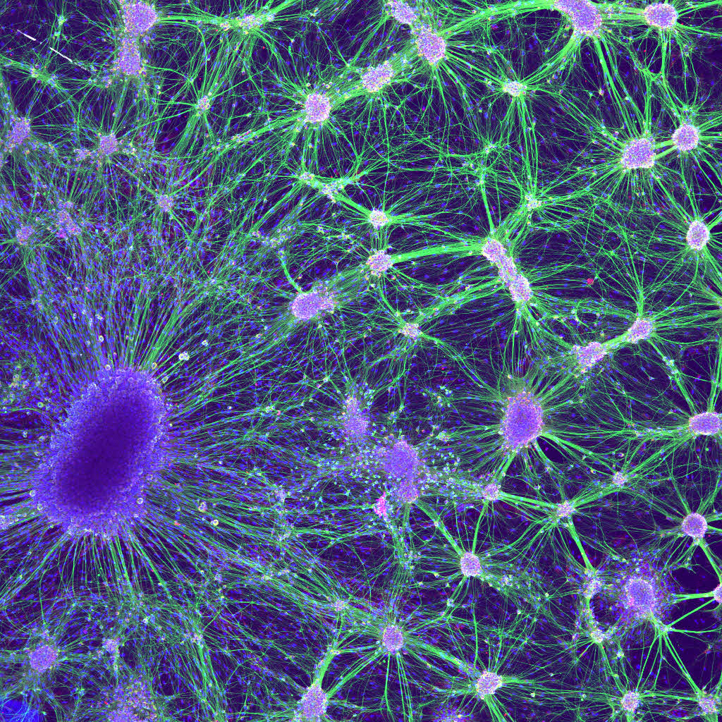
13. **Understanding Joint Innervation: How Nerves Contribute to Sensation and Stability**The intricate functionality of synovial joints is not solely dependent on their structural components but also on a sophisticated network of nerves that provide sensation and contribute to stability. Synovial joint innervation is particularly well-understood, involving both sensory and autonomic nerve fibers. The autonomic nervous system, specifically sympathetic nerves, plays a role in regulating vascular tone within the joint, influencing blood flow through interactions with smooth muscle α-1 adrenergic receptors.
Sensory nerves, found abundantly within the articular capsule and ligaments, are crucial for proprioception – our body’s sense of position and movement. Specialized receptors like Ruffini endings and Pacinian corpuscles provide this vital proprioceptive feedback. This continuous sensory input allows for the reflex control of posture, locomotion, and coordinated movement. Additionally, free nerve endings are present to convey diffuse, often poorly localized, pain sensations when a joint is strained. It’s important to note that articular cartilage itself, being avascular, has no direct nerve supply.
Two fundamental principles govern synovial joint innervation, offering insights into their integrated function. The first is the Hilton law, which states that the articular nerves supplying a joint are typically branches of the same nerves that supply the muscles responsible for moving that joint. These nerves also supply the overlying skin. This interconnectedness explains why irritation of an articular nerve can trigger a reflex muscular spasm, positioning the joint for comfort, and how joint pain can sometimes be “referred” to the skin surface.
The second principle, known as the Gardner observation, complements the Hilton law by describing a relationship vital for joint stabilization. It posits that the segment of the articular capsule that is tightened by muscle contraction is supplied by the same nerves that innervate the antagonist muscles. This arrangement facilitates local reflex arcs, providing a dynamic feedback system that enhances the stability of the joint during movement and at rest.
14. **The Dynamic Support System: Muscles and Tendons as Joint Stabilizers**While ligaments provide passive structural stability to joints, the dynamic support system of muscles and their associated tendons plays an equally, if not more, critical role in maintaining joint integrity and function, particularly in synovial joints. These muscular structures are not merely movers of bones; they are active, adaptable stabilizers that constantly respond to internal and external forces.
Muscles and their tendons span across joints, creating a robust framework that resists undesirable forces. In this capacity, they behave much like “dynamic ligaments,” offering continuous, responsive support. When muscles contract, they not only generate movement but also actively pull bones together, increasing joint compression and reinforcing its structure against dislocation or excessive strain.
The direct correlation between muscle strength and joint stability cannot be overstated. A strong musculature surrounding a joint enhances its ability to withstand stress, absorb shock, and maintain proper alignment throughout a range of movements. This dynamic stabilization is especially pronounced in synovial joints, which, due to their inherent freedom of movement, rely heavily on muscular action for both mobility and protection from injury.
As we’ve journeyed through the intricate world of human joints, from their fundamental components to their diverse classifications, clinical challenges, and sophisticated support systems, it becomes clear that these biological marvels are far more than simple connection points. They are vital for every step, every gesture, and every interaction with our environment. By understanding their complex mechanics and the factors that influence their health, we are better equipped to proactively address stiffness, mitigate pain, and ultimately nurture our body’s incredible capacity for movement and resilience. This foundational knowledge empowers us to seek effective interventions, adopt preventive measures, and embrace a lifestyle that supports lasting joint health, ensuring our bodies remain in optimal working order for years to come.


