

Marfan syndrome, a pervasive and often misunderstood genetic disorder, represents a critical area of focus within modern medicine due to its wide-ranging impact on the human body. As an inherited condition, it fundamentally compromises connective tissue – the essential fibers providing support and anchorage to organs and various bodily structures. The consequences of this systemic defect are profound, manifesting in an array of symptoms that can range from subtly mild to acutely life-threatening, touching upon the heart, eyes, blood vessels, and skeletal framework with particular intensity.
The complexity of Marfan syndrome lies not only in its diverse clinical presentation but also in its potential for severe, often unpredictable, complications. For many years, the condition posed formidable challenges to both diagnosis and management, frequently leading to diminished quality of life and shortened life expectancies. However, significant strides in medical research and clinical care have transformed the outlook for individuals affected by this disorder, offering new avenues for early detection, effective treatment, and improved long-term outcomes.
This in-depth exploration will navigate the intricacies of Marfan syndrome, starting with its foundational definition and moving through its most common and critical manifestations across various physiological systems. We will examine the distinctive physical characteristics, delve into the perilous cardiovascular complications, shed light on the array of ocular and other systemic issues, and finally, trace the condition back to its genetic roots, providing a comprehensive understanding of this remarkable and challenging disorder.
1. **Understanding Marfan Syndrome: A Multi-Systemic Connective Tissue Disorder**Marfan syndrome is fundamentally an inherited disorder, defined by a defect in the connective tissue that serves as the crucial architectural scaffolding for the body. This omnipresent tissue, composed of fibers that provide support and anchor organs and other structures, is weakened and compromised throughout the body in individuals with Marfan syndrome. The pervasive nature of connective tissue means that the disorder can impact almost any part of the body, leading to a highly variable and often complex clinical picture, even among members of the same family.
The primary systems most commonly affected by Marfan syndrome include the heart and blood vessels, the eyes, and the skeleton. However, its influence extends beyond these core areas, frequently involving the lungs and the nervous system as well. The spectrum of damage caused by the syndrome is remarkably broad, ranging from manifestations so mild they might go unnoticed for years, to severe, rapidly progressing conditions that demand immediate and intensive medical intervention. This variability underscores the importance of a holistic and individualized approach to diagnosis and ongoing care.
At its core, the condition is rooted in a genetic change that impairs the body’s ability to produce a healthy, functional protein vital for the elasticity and strength of connective tissue. This foundational defect, although singular in its origin, precipitates a cascade of effects that can compromise the integrity and function of numerous bodily systems. Understanding this fundamental mechanism is key to appreciating the diverse symptoms and complications that characterize Marfan syndrome and the rationale behind its multifaceted management strategies.
2. **The Distinctive Physical Signature: Skeletal Manifestations**One of the most recognizable aspects of Marfan syndrome lies in its distinctive impact on the skeletal system, which often provides the earliest visible clues to the condition. Individuals with Marfan syndrome are typically described as tall and thin, frequently presenting with unusually long arms, legs, fingers, and toes – a characteristic often referred to as arachnodactyly. These disproportionate limb lengths can be striking, with an arm span often exceeding a person’s height, prompting specific measurements by medical professionals during diagnostic evaluations.
Beyond mere height and limb proportions, the skeletal system exhibits a range of specific deformities and signs. Many individuals may develop an abnormally curved spine, a condition known as scoliosis, or other spinal abnormalities such as kyphosis. The breastbone, or sternum, is also commonly affected, either protruding outward (pectus carinatum) or dipping inward (pectus excavatum), interfering with normal rib development. Other frequently observed features include a high, arched palate in the mouth often accompanied by crowded teeth, flat feet (pes planus), and joints that exhibit unusual flexibility or hypermobility, making them prone to dislocation.
Clinical examination includes specific tests to identify these characteristic skeletal features. The “thumb sign,” also known as Steinberg’s sign, is positive when the tip of the thumb extends far beyond the edge of the palm when a fist is made. Similarly, the “wrist sign,” or Walker-Murdoch sign, is positive if the little finger and thumb overlap when curled around the opposite wrist, reflecting unusually long fingers and thin wrists. These signs, while not definitive on their own, serve as important indicators that guide clinicians toward further investigation and a potential diagnosis of Marfan syndrome, highlighting the pervasive nature of the connective tissue defect in shaping the body’s architecture.

3. **Cardiovascular Vulnerabilities: The Aorta and Heart Valves**The most perilous and often life-threatening complications associated with Marfan syndrome invariably involve the cardiovascular system, particularly the aorta and the heart’s delicate valves. Faulty connective tissue critically weakens the aorta, the body’s largest artery that emerges directly from the heart and functions as the primary conduit for oxygenated blood to the entire body. This inherent structural fragility makes the aorta susceptible to a range of serious issues that can rapidly escalate into medical emergencies.
A primary concern is an aortic aneurysm, a condition where the pressure of blood exiting the heart causes the wall of the aorta to bulge outward, akin to a weak spot in a tire. In individuals with Marfan syndrome, this phenomenon is most frequently observed at the aortic root, the segment of the artery closest to the heart. Such enlargement significantly increases the risk of an aortic dissection – a catastrophic event where a small tear in the innermost layer of the aortic wall allows blood to penetrate and separate the inner and outer layers. This dissection can cause excruciating, tearing pain in the chest or back and, if left untreated, can lead to a fatal rupture of the vessel.
Beyond the aorta itself, the heart valves are also highly vulnerable due to the compromised connective tissue. Conditions such as mitral valve prolapse are common, where the flaps of the mitral valve, controlling blood flow on the left side of the heart, become “floppy” and fail to close tightly. This can lead to blood leaking back into the heart (regurgitation), forcing the heart to work harder and potentially leading to heart failure over time. Similarly, aortic regurgitation can occur when the aortic valve does not fully close, allowing blood to leak back into the heart. These cardiovascular issues, from aneurysms to valve malformations, necessitate vigilant monitoring and often require proactive medical or surgical intervention to mitigate the severe risks they pose.

4. **Ocular Complications: Beyond Vision Impairment**The eyes, too, are profoundly affected by the systemic connective tissue defect inherent in Marfan syndrome, presenting a unique set of challenges that extend beyond simple vision impairment. The most distinctive ocular manifestation is partial lens dislocation, medically termed ectopia lentis. This occurs in more than half of those with the syndrome, where the focusing lens within the eye shifts out of its normal position due to weakness in the ciliary zonules – the delicate connective tissue strands responsible for suspending the lens. The mutations responsible for Marfan syndrome directly weaken these zonules, causing them to stretch and allow the lens to displace, most commonly upwards and outwards.
This dislocation, even if subtle, can lead to significant visual disturbances, including extreme nearsightedness (myopia) or blurred vision, depending on the degree and direction of the lens shift. While partial lens dislocation is a hallmark, individuals with Marfan syndrome are also at a heightened risk for several other serious eye conditions. These include retinal detachment or tears, where the light-sensitive tissue lining the back of the eye pulls away from its supportive layer, potentially leading to permanent vision loss if not promptly treated.
Furthermore, these individuals tend to develop eye problems typically seen in older populations at a much younger age. Early-onset glaucoma, characterized by increased pressure within the eye that can damage the optic nerve, and cataracts, which are cloudy areas in the eye’s normally clear lens, are significant concerns. Given the fragility of ocular structures, particularly the retina, those diagnosed with Marfan syndrome are often advised to avoid contact sports and other activities that carry a risk of falls or direct impact to the head, emphasizing the comprehensive care required to preserve vision and prevent further complications.

5. **Pulmonary and Neurological Challenges: Hidden Impacts**While Marfan syndrome is widely recognized for its effects on the skeletal, cardiovascular, and ocular systems, its pervasive influence extends to other critical areas of the body, including the lungs and the nervous system. These manifestations, though perhaps less frequently highlighted than cardiac issues, can significantly impact an individual’s quality of life and, in some instances, pose serious health risks. Understanding these broader systemic impacts is essential for a complete clinical picture and effective management.
In the pulmonary system, individuals with Marfan syndrome may experience various lung-related problems. One notable complication is spontaneous pneumothorax, where air escapes from a lung and occupies the pleural space between the chest wall and the lung, causing it to partially compress or collapse. This can lead to symptoms such as pain, shortness of breath, and, if severe, cyanosis, requiring immediate medical attention. Other potential pulmonary issues include sleep apnea, often managed with respiratory support, and idiopathic obstructive lung disease. Pathologic changes in lung tissue, such as cystic changes, emphysema, pneumonia, and bronchiectasis, have also been described, underscoring the broad impact on respiratory health.
The nervous system, specifically the spinal cord and its coverings, can also be affected, leading to a condition known as dural ectasia. This involves the weakening of the connective tissue of the dural sac, which encases the spinal cord. Dural ectasia can exist asymptomatically for an extended period but may eventually manifest as lower back pain, leg pain, abdominal discomfort, other neurological symptoms in the lower extremities, or headaches that often improve when lying flat. While not always visible in early stages on X-rays, a worsening of symptoms typically prompts an MRI of the lower spine, revealing a dilated pouch that can wear away at the lumbar vertebrae, illustrating another profound, yet often hidden, systemic impact of Marfan syndrome.

6. **The Genetic Underpinning: FBN1 Mutation and Autosomal Dominant Inheritance**At the very foundation of Marfan syndrome lies a specific genetic defect: a mutation in the *FBN1* gene, located on chromosome 15. This gene is responsible for providing the instructions for the body to produce fibrillin-1, a crucial glycoprotein component of the extracellular matrix. Fibrillin-1 is indispensable not only for the structural integrity of connective tissue but also for its elasticity and strength. It plays a pivotal role in the biogenesis and maintenance of elastic fibers, which are abundantly found in vital structures such as the aorta, ligaments, and the ciliary zonules of the eye—areas that are consequently among the most severely affected in Marfan syndrome.
The inheritance pattern of Marfan syndrome is autosomal dominant, meaning that an individual only needs to inherit one copy of the altered *FBN1* gene from a parent to develop the condition. This inheritance mechanism dictates that each child of an affected parent faces a 50 percent chance of inheriting the defective gene. In the majority of cases, approximately 75 percent, the syndrome is inherited from a biological parent who also has the disorder, leading to a traceable family history. However, in a significant minority of cases, about 25 percent, the abnormal gene arises from a *de novo* genetic mutation, meaning it develops spontaneously in the affected individual without being inherited from either parent.
Beyond its direct structural role, fibrillin-1 also serves a vital regulatory function by directly binding a latent form of transforming growth factor beta (TGF-β), thereby sequestering it and preventing it from exerting its biological activity. In Marfan syndrome, reduced levels of functional fibrillin-1 are thought to allow TGF-β levels to rise due to inadequate sequestration. While the precise mechanisms by which elevated TGF-β levels contribute to the disease’s specific pathology are still being elucidated, it is understood to trigger an inflammatory reaction that degrades elastic fibers and other components of the extracellular matrix. This pathway highlights the intricate molecular underpinnings of Marfan syndrome and its complex pathogenesis, moving beyond a simple structural defect to involve critical cellular signaling.

7. **Precise Diagnostic Methodologies: The Ghent Criteria and Beyond**Diagnosing Marfan syndrome often presents a complex challenge, particularly in children who may not exhibit clear symptoms until they reach pubescence. To standardize this critical process, international agreements have established specific diagnostic criteria, with initial guidelines agreed upon in 1996. These criteria typically involve assessing family history in conjunction with a combination of major and minor indicators of the disorder that are rare in the general population, such as four skeletal signs alongside one or more signs in other body systems like ocular and cardiovascular.
The diagnostic landscape evolved significantly with the introduction of the Revised Ghent Nosology in 2010, which superseded previous agreements and provided updated criteria. This revised framework outlines seven distinct pathways that can lead to a diagnosis, simplifying the process based on the presence or absence of a family history of Marfan syndrome. For individuals without a known family history, a diagnosis might be confirmed by a combination of factors, including a significant aortic root Z-score, ectopia lentis, or an *FBN1* mutation.
Key to these revised criteria is a comprehensive systemic score, which assigns points for various manifestations. For instance, both the wrist and thumb signs together contribute three points, while either sign alone yields one. Pectus carinatum deformity adds two points, in contrast to pectus excavatum or chest asymmetry, which provide one. Other indicators contributing to this score include hindfoot deformity, dural ectasia, protrusio acetabuli, pneumothorax, specific facial features, and even seemingly minor signs such as skin striae or myopia greater than three diopters.
Specific clinical examinations are crucial for identifying these systemic features. The “thumb sign,” also known as Steinberg’s sign, is elicited by asking the person to flex the thumb as far as possible and then close the fingers over it. A positive sign is observed when the entire distal phalanx is visible beyond the ulnar border of the hand, indicating both hypermobility and an unusually long thumb. Similarly, the “wrist sign,” or Walker-Murdoch sign, is positive if the little finger and thumb overlap when curled around the opposite wrist, reflecting unusually long fingers and thin wrists.
Beyond these physical signs and scoring systems, modern diagnostic approaches frequently integrate genetic testing through DNA analysis to identify mutations in the *FBN1* gene. Imaging techniques, such as ultrasound of the aortic root, are also vital for detecting the characteristic dilation. This multi-pronged approach ensures a precise and comprehensive diagnosis, guiding timely intervention and management strategies for individuals with Marfan syndrome.

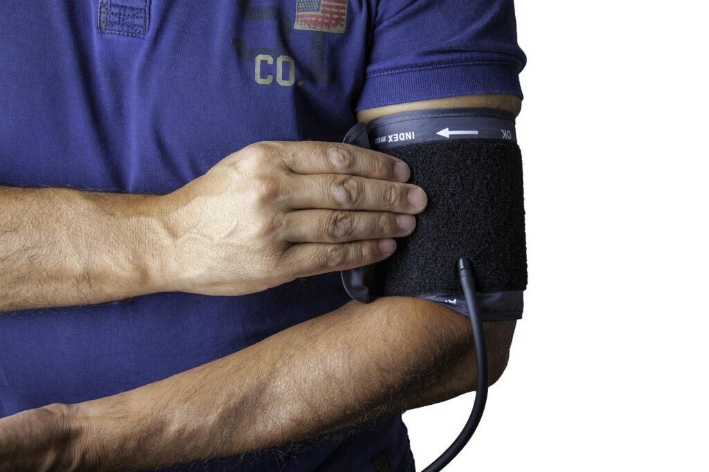
8. **Pharmacological Management: Stabilizing the Cardiovascular System**While a definitive cure for Marfan syndrome remains elusive, significant advancements in medical management have dramatically improved life expectancy, now often comparable to that of the general population. The cornerstone of treatment involves proactive medication strategies, even for young children, aimed at slowing the progression of aortic dilation and preventing damage to the heart valves. This involves meticulously controlling the individual’s heart rate, minimizing strain on the aorta, and maintaining optimal blood pressure levels.
The primary class of medications employed in this strategy includes beta blockers, such as propranolol and atenolol. These agents work by reducing the stress exerted on the aorta, thereby helping to decrease the rate of aortic dilation. By lowering the heart rate and blood pressure, beta blockers mitigate the forces that can weaken the aortic wall, which is particularly vulnerable in individuals with compromised connective tissue. Their consistent use is a critical component of preventing severe cardiovascular complications.
For individuals who may not tolerate beta blockers, alternative pharmacological options are available. Calcium channel blockers can be utilized, providing similar benefits in managing blood pressure and reducing cardiac workload. Additionally, ACE inhibitors and angiotensin receptor blockers (ARBs) are important components of a comprehensive treatment regimen. These medications contribute to lowering blood pressure and may offer protective effects on the cardiovascular system, working in conjunction with beta blockers or as alternatives.
Regardless of the specific medication regimen, regular checkups are an indispensable part of managing Marfan syndrome. These appointments allow medical professionals to monitor the health of the heart valves and the aorta meticulously, ensuring that the chosen pharmacological interventions are effective and adjusted as necessary. This vigilant monitoring helps to preemptively address any progression of the condition and maintain cardiovascular stability, underscoring the long-term commitment required for optimal care.
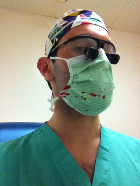
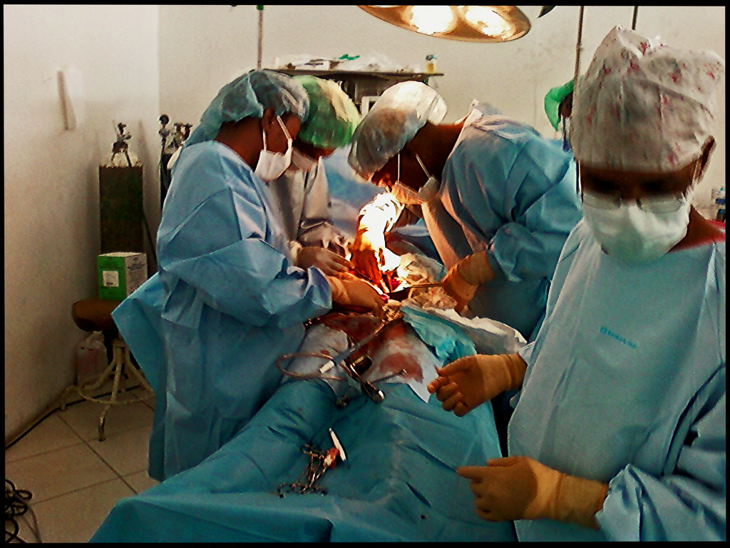
9. **Surgical Interventions: Repairing and Protecting the Aorta**When the dilation of the aorta reaches a significant diameter, or if a dissection, rupture, or severe heart valve failure occurs, surgical intervention becomes a necessary and often life-saving measure for individuals with Marfan syndrome. Elective aortic graft surgery, while a serious undertaking, generally boasts high success rates, especially when performed proactively before a critical event. This planned approach allows for detailed preparation and reduces the risks associated with emergency procedures.
Specific surgical procedures commonly employed include the composite aortic valve graft or valve-sparing aortic root replacement. These operations are typically considered when the aortic root diameter reaches approximately 50 millimeters, though each case requires a personalized evaluation by a qualified cardiologist. The goal is to reinforce or replace the weakened section of the aorta, preventing catastrophic events like dissection or rupture. Newer valve-sparing techniques are also becoming increasingly common, aiming to preserve the patient’s native heart valves when feasible.
It is crucial to differentiate between elective surgery and emergency procedures following acute aortic dissection or rupture. The latter scenarios present considerably greater challenges and higher risks. As individuals with Marfan syndrome survive initial aortic root enlargements and dissections of the ascending aorta, other vascular repairs, such as those for descending thoracic aortic aneurysms or aneurysms in other vessels like the subclavian arteries, are becoming more frequently performed, highlighting the evolving nature of long-term care.
Beyond cardiovascular issues, the skeletal and ocular manifestations of Marfan syndrome may also necessitate surgical or interventional treatments, although these are typically not life-threatening. Skeletal symptoms can be managed with pain medications or muscle relaxants, but surgical correction may be required for severe spinal abnormalities or chest deformities. Any spinal surgery for a person with Marfan syndrome demands detailed imaging and meticulous planning due to potential asymptomatic spinal abnormalities.
For ocular complications, surgery can often restore or preserve vision. Ectopia lentis, the partial dislocation of the eye lens, can be addressed through artificial lens implantation. Furthermore, surgical interventions are available to treat glaucoma, repair retinal detachments or tears, and remove cataracts, all of which are conditions that individuals with Marfan syndrome tend to develop at an earlier age. In cases of spontaneous pneumothorax, treatment varies based on severity, from watchful waiting for small collapses to chest drain management or emergency decompression for larger, more critical events. Custom-built supports for the aortic root are also being explored as an alternative therapeutic approach.

10. **Physical Activity Guidelines: Balancing Health and Risk**Given the systemic nature of Marfan syndrome and its profound impact on connective tissue, particularly within the cardiovascular system, careful consideration of physical activity is paramount. Individuals with the condition are generally advised to avoid strenuous exercise to minimize strain on the aorta and other vulnerable structures. The American Heart Association (AHA) has issued specific recommendations to guide patients and clinicians in making informed decisions about safe levels of activity.
The AHA identifies a range of activities generally considered permissible for individuals with Marfan syndrome who exhibit no or only mild aortic dilation. These include recreational pursuits such as bowling, golf, and ice skating (though not ice hockey due to its higher intensity). Other safe options encompass snorkeling, brisk walking, using a treadmill, stationary biking, modest hiking, and participating in doubles or singles tennis. These activities allow for physical conditioning without unduly elevating cardiovascular risk.
Intermediate-risk activities represent a category where individuals need to exercise greater caution and potentially seek more specific medical guidance. This group includes sports like basketball, racquetball, and squash, as well as running (both sprinting and jogging) and various forms of skiing. Other activities falling into this category are soccer, touch (flag) football, baseball, softball, biking, lap swimming, motorcycling, and horseback riding. The decision to engage in these activities should always involve a thorough discussion with a healthcare provider, weighing the individual’s specific cardiovascular status and overall health.
Conversely, certain high-risk activities are strongly discouraged due to the significant strain they place on the cardiovascular and musculoskeletal systems. These include highly intense forms of exercise such as bodybuilding and weightlifting, encompassing both non-free and free weights. Contact sports like ice hockey and activities with a high potential for falls or sudden impacts, such as rock climbing, windsurfing, surfing, and scuba diving, are also generally advised against. Adherence to these guidelines is crucial for mitigating the risk of serious complications like aortic dissection or rupture, thereby supporting long-term health and well-being.

11. **Differential Diagnosis: Distinguishing Marfan from Mimics**One of the persistent challenges in diagnosing Marfan syndrome lies in its clinical overlap with several other genetic disorders that can produce similar physical characteristics, often referred to as a “marfanoid” habitus. Distinguishing Marfan syndrome from these mimicking conditions is critical for accurate diagnosis and appropriate management. The process relies heavily on a combination of thorough clinical evaluation, detailed family history, and, increasingly, advanced genetic testing to pinpoint specific underlying mutations.
Several conditions present with features that can be confused with Marfan syndrome. Congenital contractural arachnodactyly, also known as Beals–Hecht syndrome, shares traits such as long limbs and joint contractures. Ehlers–Danlos syndrome, a group of disorders affecting connective tissue, can manifest with joint hypermobility and skin elasticity that might initially suggest Marfan syndrome. Another important differential is homocystinuria, an inherited disorder of amino acid metabolism, which can also lead to skeletal abnormalities, ectopia lentis, and vascular issues.
The list of marfanoid conditions extends further to include Loeys–Dietz syndrome, a relatively newer diagnosis that shares significant clinical overlap with Marfan syndrome, particularly concerning aortic root dilation. Differentiating between these two is especially important because Loeys–Dietz syndrome is associated with mutations in genes encoding transforming growth factor beta (TGF-β) receptors, leading to different management considerations. Other conditions such as the MASS phenotype (Mitral valve prolapse, Aortic enlargement, Skin striae, Skeletal features), Multiple endocrine neoplasia type 2B, Shprintzen–Goldberg syndrome, and Stickler syndrome also warrant consideration in a differential diagnosis.
Given the nuanced similarities and critical differences between these conditions, genetic testing plays an indispensable role. While clinical criteria, like the revised Ghent nosology, guide the initial assessment, definitive diagnosis often requires DNA analysis to identify specific gene mutations. For example, understanding the involvement of the TGFβR2 gene in Loeys–Dietz syndrome, as opposed to *FBN1* in Marfan, underscores the importance of precise genetic identification to ensure patients receive the most appropriate and targeted medical care, addressing their unique pathophysiological mechanisms.

12. **Prognosis and Pregnancy: Navigating Life with Marfan Syndrome**The prognosis for individuals with Marfan syndrome has dramatically improved since the 1970s, largely due to advancements in diagnostic tools and comprehensive treatment strategies. While there is no known cure for the underlying genetic defect, many individuals with proper medical management can now achieve a life expectancy similar to that of the average person. This positive shift underscores the importance of early diagnosis, vigilant monitoring, and adherence to prescribed treatments.
A particularly critical area of concern for individuals with Marfan syndrome is pregnancy, which introduces unique and potentially life-threatening risks. The condition inherently weakens the walls of the aorta, the body’s main artery. During pregnancy, the cardiovascular system undergoes significant changes, with the heart pumping a considerably larger volume of blood than usual to support both the mother and the developing fetus. This increased workload places immense additional stress on an already compromised aorta.
The heightened strain on the aortic wall during pregnancy significantly elevates the risk of a deadly aortic dissection or rupture. These catastrophic events can occur suddenly and have severe consequences for both the mother and the baby. Consequently, pregnancy in individuals with Marfan syndrome necessitates exceptionally vigilant medical management, often involving a multidisciplinary team of specialists including cardiologists, geneticists, and high-risk obstetricians, to monitor cardiovascular health closely and intervene swiftly if complications arise.
Navigating life with Marfan syndrome is a journey that requires ongoing medical attention, proactive management, and a deep understanding of the condition’s potential impacts across various bodily systems. From meticulous diagnostic methodologies and a tailored pharmacological regimen to potential surgical interventions, lifestyle adjustments, and specialized care during critical life stages like pregnancy, comprehensive and individualized care is paramount. With continued research and dedicated healthcare teams, individuals living with Marfan syndrome can look forward to maintaining a high quality of life and improved long-term outcomes, embracing a future that was once considered unimaginable.
In essence, Marfan syndrome, while complex and challenging, is a condition where informed vigilance and proactive medical care can make all the difference. The ongoing advancements in diagnostics, treatments, and understanding of its intricate genetic and physiological underpinnings offer a beacon of hope, transforming the lives of those affected and ensuring they can lead full, productive lives. The journey with Marfan syndrome is a testament to the power of modern medicine and the resilience of the human spirit.


