

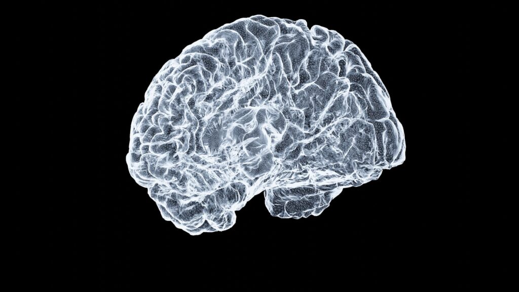
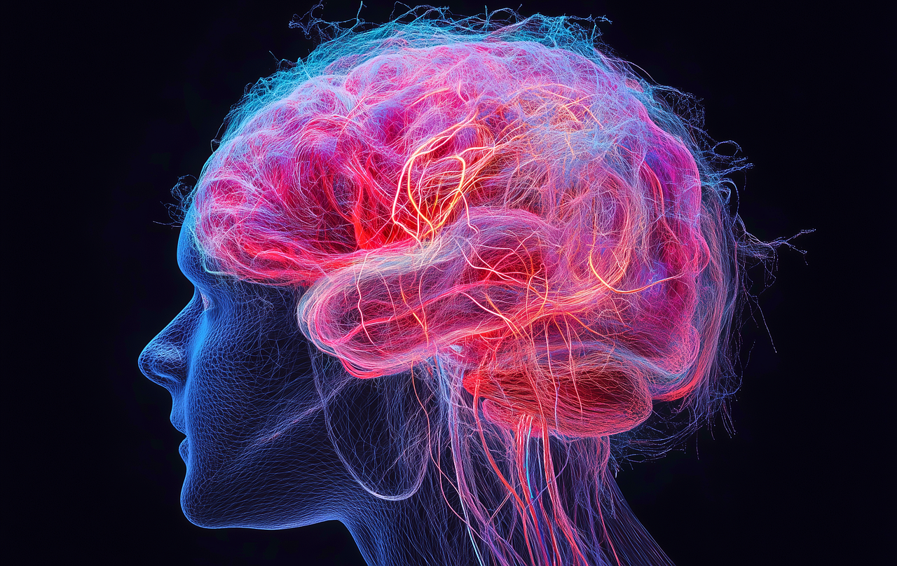
The human brain, a masterpiece of biological engineering, stands as the body’s most complex organ. It orchestrates thought, memory, emotion, and all physiological processes essential for life. Far from a simple mass, it is a highly organized structure: the average adult brain weighs about 3 pounds, comprising 60% fat and 40% water, protein, carbohydrates, and salts. Crucially, it is not a muscle but a vibrant network of blood vessels, nerves, billions of neurons, and supporting glial cells. Together with the spinal cord, it forms the central nervous system (CNS)—the body’s master control unit. Its core function lies in sending and receiving chemical and electrical signals: some regulate tiredness or pain, others travel via the spinal cord to extremities, enabling instantaneous communication that governs all interactions with the world.
Gray Matter and White Matter: The Brain’s Functional Tissues
The CNS contains two primary tissue types: gray matter and white matter, distinct in composition and role. In the brain, gray matter forms the darker outer layer (cerebral cortex), while white matter occupies the lighter inner region. In the spinal cord, this arrangement inverts—white matter on the outside, gray matter inside.
Gray matter consists mainly of neuron somas (cell bodies), dendrites, and synapses, critical for receiving and processing information. It handles muscle control, sensory perception, memory, emotion, and speech. White matter, by contrast, comprises axons (long neuron stems) wrapped in myelin—a fatty, protective coating that gives it a light color and speeds electrical signal transmission. White matter acts as a conduit, relaying processed information across the nervous system, much like cables connecting computer components.
Major Brain Structures: Cerebrum, Brainstem, and Cerebellum
At a macroscopic level, the brain divides into three key parts, each with unique roles.
The Cerebrum: Seat of Higher Cognition
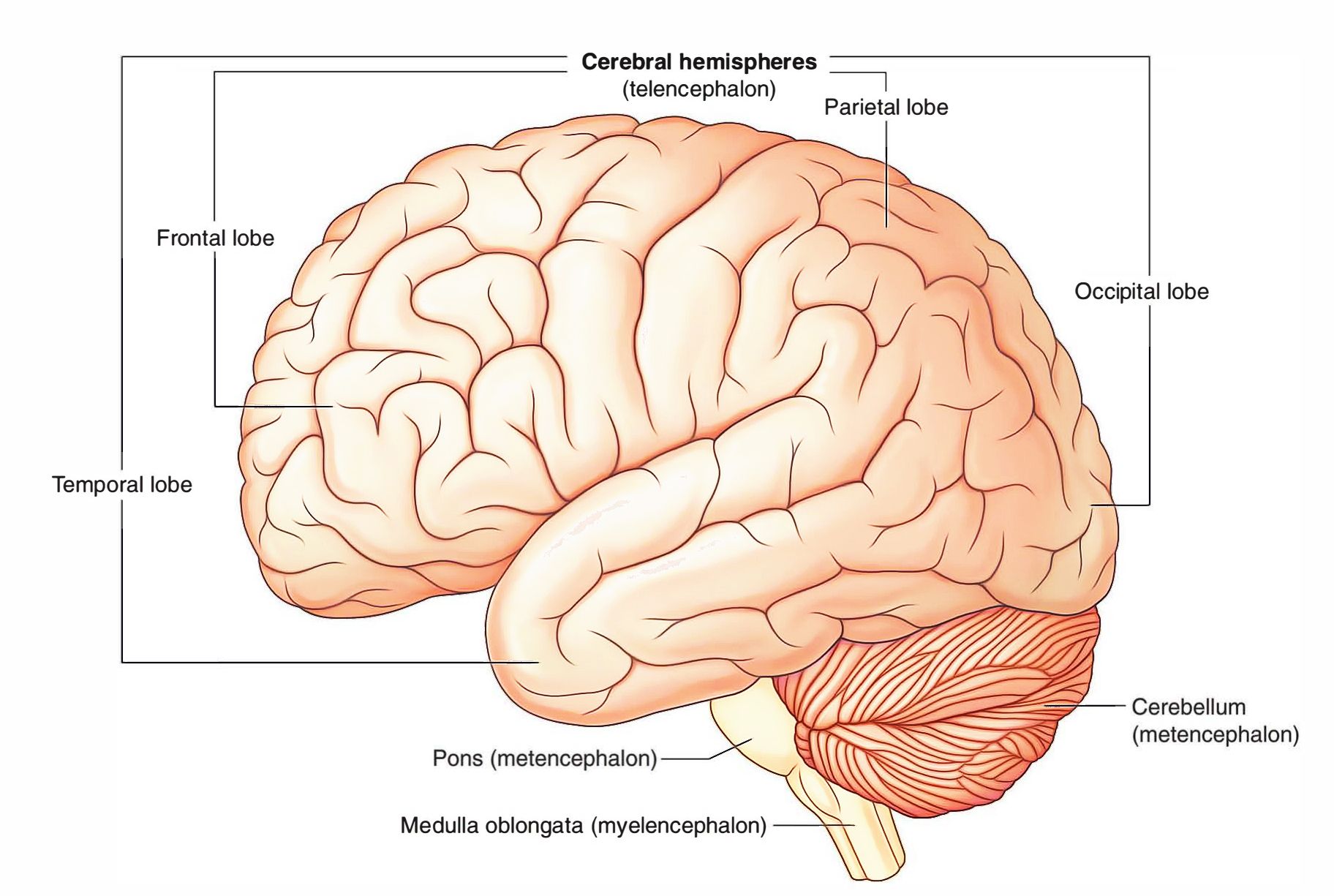
The largest brain structure, the cerebrum occupies the upper cranial cavity and governs higher cognitive functions. It initiates voluntary movements, regulates body temperature, and enables speech, judgment, reasoning, problem-solving, emotion, and learning. It also processes sensory input from vision, hearing, touch, taste, and smell, integrating these to form a coherent perception of the world.
The cerebrum includes gray matter (cerebral cortex) and inner white matter. Its cortex—named for its “bark-like” outer layer—features ridges (gyri) and folds (sulci) that increase surface area, enhancing processing capacity. It makes up about half the brain’s weight. The cortex splits into left and right hemispheres, separated by the interhemispheric fissure. In a phenomenon called contralateral control, the right hemisphere manages the left body, and the left hemisphere controls the right. The corpus callosum—a thick C-shaped bundle of white matter—connects the hemispheres, ensuring seamless information integration.
The Brainstem: Vital for Survival
Beneath the cerebrum, the brainstem links the cerebrum to the spinal cord, regulating automatic, life-sustaining functions. It has three parts:
- Midbrain (Mesencephalon): A complex structure with neuron clusters, nuclei, and pathways. It processes auditory information, coordinates movement (including eye movements), and includes the substantia nigra—rich in dopamine neurons, critical for movement and affected in Parkinson’s disease.
- Pons: Connects the midbrain to the medulla. It houses four cranial nerves, controlling functions like tear production, chewing, blinking, vision focus, balance, hearing, facial expressions, swallowing, bladder function, posture, and sleep.
- Medulla Oblongata: Located at the brainstem’s base, merging with the spinal cord. It regulates heart rhythm, breathing, blood flow, oxygen/carbon dioxide levels, and reflexes like sneezing, vomiting, coughing, and swallowing.
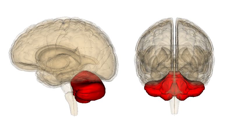
The Cerebellum: Coordination and Beyond
Nicknamed the “little brain,” the cerebellum sits at the back of the head, beneath the temporal and occipital lobes. It has two hemispheres, with an outer layer of neurons and inner connections to the cerebral cortex. Its primary role is coordinating smooth, precise voluntary movements, maintaining posture, balance, and equilibrium. Emerging research suggests it may also influence higher functions like thought, emotions, social behavior, and be linked to conditions such as addiction, autism, and schizophrenia.
Protecting the Brain: A Multi-Layered Defense
The brain is shielded by several protective layers:
- Cranium: The skull’s bony structure forms the first physical barrier against external forces.
- Meninges: Three specialized layers surrounding the brain and spinal cord:
- Dura Mater: A thick, tough outer layer lining the skull, with two sublayers (periosteal and meningeal) that house blood vessels for brain circulation.
- Arachnoid Mater: A thin, weblike layer without nerves or blood vessels. Below it lies the subarachnoid space, filled with cerebrospinal fluid (CSF)—a clear fluid cushioning the brain and spinal cord, removing waste, and delivering nutrients.
- Pia Mater: A delicate inner layer clinging to the brain’s surface (following gyri and sulci), rich in blood vessels supplying nutrients to brain tissue.
Blood Supply: Sustaining the Brain’s Activity
The brain’s constant activity demands uninterrupted blood and oxygen. Two primary vessel sets deliver this:
- Carotid Arteries: External carotids supply the head and face; internal carotids enter the skull, nourishing the front brain (critical for thought, memory, speech).
- Vertebral Arteries: Ascend the spinal column, merging at the brainstem to form the basilar artery, which supplies the rear brain (cerebellum, brainstem) for balance, breathing, and heart rate.
The circle of Willis—a loop of vessels near the brain’s base—connects these arteries, allowing collateral blood flow. This redundancy protects against flow interruptions, ensuring the brain’s metabolic needs are met.
Cerebral Lobes: Specialized Functional Zones
The cerebrum’s cortex divides into four lobes, each with distinct roles:
- Frontal Lobe: Behind the forehead, the largest lobe governs executive functions (personality, decision-making, planning), voluntary movements, smell recognition, and speech production (via Broca’s area). Damage impairs speaking ability.
- Parietal Lobe: Behind the frontal lobe, processes sensory information (touch, temperature, pain), identifies objects, interprets spatial relationships, and aids language comprehension (via Wernicke’s area).
- Occipital Lobe: At the brain’s back, exclusively processes visual information—transforming light signals into coherent images, enabling object/color recognition and depth perception.
- Temporal Lobes: Near the ears, handle auditory processing (speech, music), short-term memory, smell recognition, emotion processing, and long-term memory retrieval.
Hemispheric Specialization: Left and Right Brain Roles
The cerebrum’s two hemispheres exhibit specialized processing, though they collaborate via the corpus callosum:
- Left Hemisphere: Dominant in language (reading, writing), mathematical reasoning, sequential problem-solving, grammar, and analytical thinking.
- Right Hemisphere: Specializes in spatial awareness, creativity, face recognition, emotion interpretation, pattern visualization, and musical/artistic abilities.
Complex tasks require both hemispheres: the left processes speech words, while the right interprets tone and context, enabling full communication understanding.
Subcortical Structures: Hidden Regulators
Deep brain structures fine-tune vital processes:
- Thalamus: A “relay station” above the brainstem, filtering sensory/motor signals to the cortex, regulating consciousness, sleep, and alertness (all senses except smell pass through it).
- Hypothalamus: Below the thalamus, regulates hormones (via the pituitary gland), body temperature, sleep, hunger, thirst, and influences memory/emotion.
- Pituitary Gland: A pea-sized “master gland” controlled by the hypothalamus, regulating other glands (thyroid, adrenals, ovaries, testicles) to influence growth, metabolism, reproduction, and stress.
- Amygdalae: Almond-shaped structures in the temporal lobes, key to emotion regulation (especially fear), reward processing, and the “fight or flight” response.
- Hippocampus: Seahorse-shaped in the temporal lobes, critical for forming long-term memories and spatial navigation; linked to Alzheimer’s disease.
- Basal Ganglia: Deep nuclei (including the striatum, globus pallidus) aiding smooth voluntary movements (with the substantia nigra); also influence emotions and habit formation.
- Pineal Gland: Attached to the third ventricle, secretes melatonin to regulate circadian rhythms (sleep-wake cycles).
Ventricles and CSF Circulation
The brain contains four interconnected ventricles—open areas producing CSF. CSF circulates through ventricles, the spinal canal, and the subarachnoid space, cushioning the CNS, removing waste, and delivering nutrients, maintaining a stable environment for neurons.
Cranial Nerves: Direct Brain Connections
Twelve cranial nerve pairs link the brain to the head, neck, and torso, handling sensory and motor functions:
- Olfactory Nerve (I): From the cerebrum, governs smell.
- Optic Nerve (II): From the cerebrum, transmits visual information.
- Oculomotor (III), Trochlear (IV), Abducens (VI) Nerves: From the brainstem, control eye movements and pupil response.
- Trigeminal Nerve (V): From the pons, the largest cranial nerve, conveying facial sensation and enabling chewing.
- Facial Nerve (VII): From the pons, controls facial movements, taste (front tongue), and tear production.
- Vestibulocochlear Nerve (VIII): From the pons, handles balance and hearing.
- Glossopharyngeal Nerve (IX): From the medulla, manages taste (back tongue), throat movements, and swallowing.
- Vagus Nerve (X): From the medulla, influences ear/digestive sensation, and controls heart, throat, and digestive motor functions.
- Accessory Nerve (XI): From the medulla, innervates head, neck, and shoulder muscles (e.g., shrugging).
- Hypoglossal Nerve (XII): From the medulla, controls tongue movement for speech and swallowing.
The brain’s ability to adapt—neuroplasticity—allows neurons to form new connections, reorganize pathways, and compensate for damage. This flexibility underlies learning, memory, skill acquisition, and adaptation to new environments, reflecting the brain’s dynamic nature and capacity for continuous growth.
The human brain is more than a collection of parts; it is an integrated system where each region collaborates to enable thought, emotion, and action. From its core structures to specialized networks, every component works in harmony, supported by protective mechanisms and adaptive abilities. This complexity underscores its role as a biological marvel, defining human existence.


